
Is Your Spinal Stenosis Causing Back Pain?
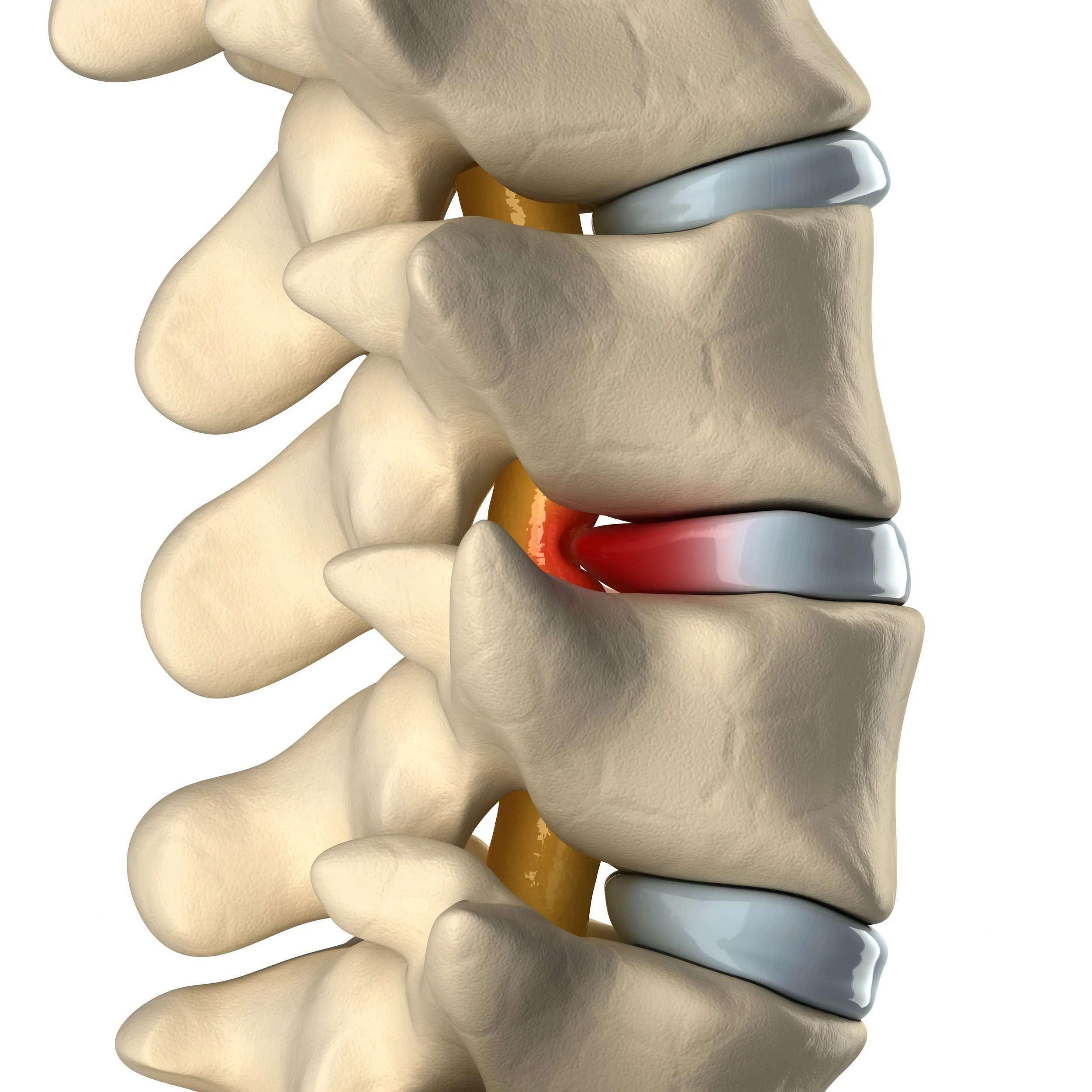
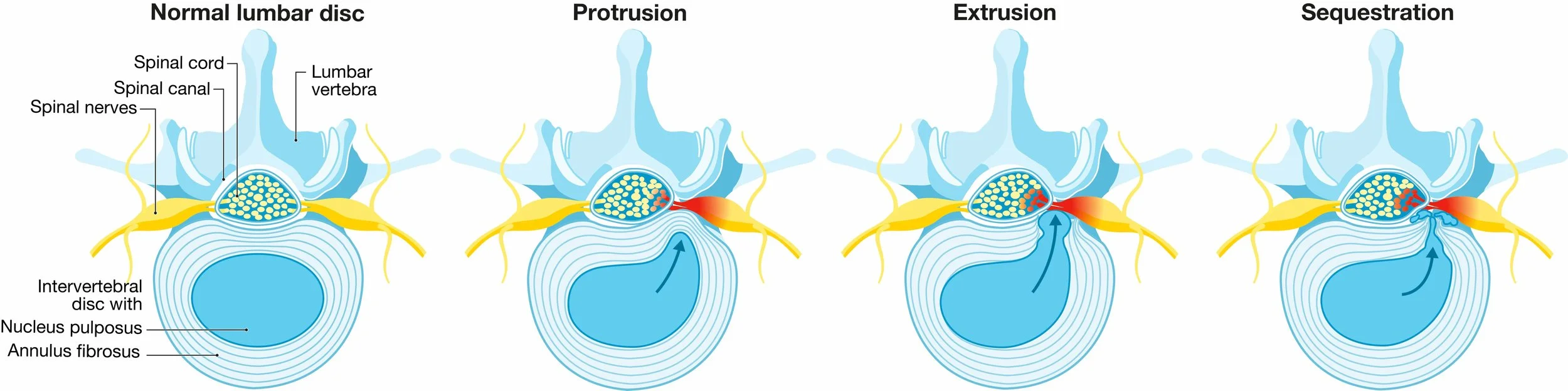
Bulging disc
SO YOU HAVE A SPINAL STENOSIS DIAGNOSIS…
In this post I will cover:
The anatomy
What causes Spinal Stenosis
Maybe Spinal Stenosis is not causing your back pain
o Scans have limitations
o Back muscle damage
o Misaligned pelvis causing tilted vertebrae
Case: Teen with Chronic Back Pain
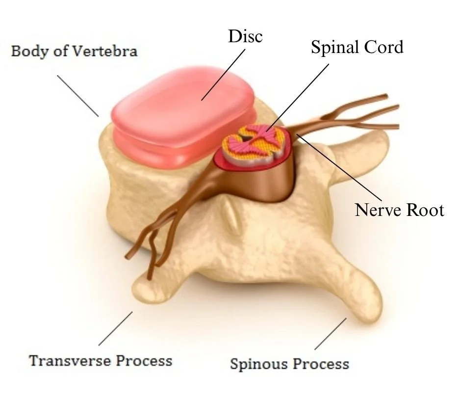
Anatomy
The spine consists of:
A stacked column of 24 bones called vertebra;
The spinal cord inside those protective vertebrae, extending from the brain at the base of the skull to the sacrum; and,
Nerves that exit the spinal column. Those sensory and motor nerves feed every part of the body. If those nerves are damaged, you’re in pain, you may have numbness or tingling in a body part, or worst case, a part of the body does not work at all; e.g., paraplegic.
What Causes Spinal Stenosis
When the space around the spinal cord encased by the column of stacked boney vertebrae is narrower than it should be, there can be pressure on the spinal cord and nerve roots.
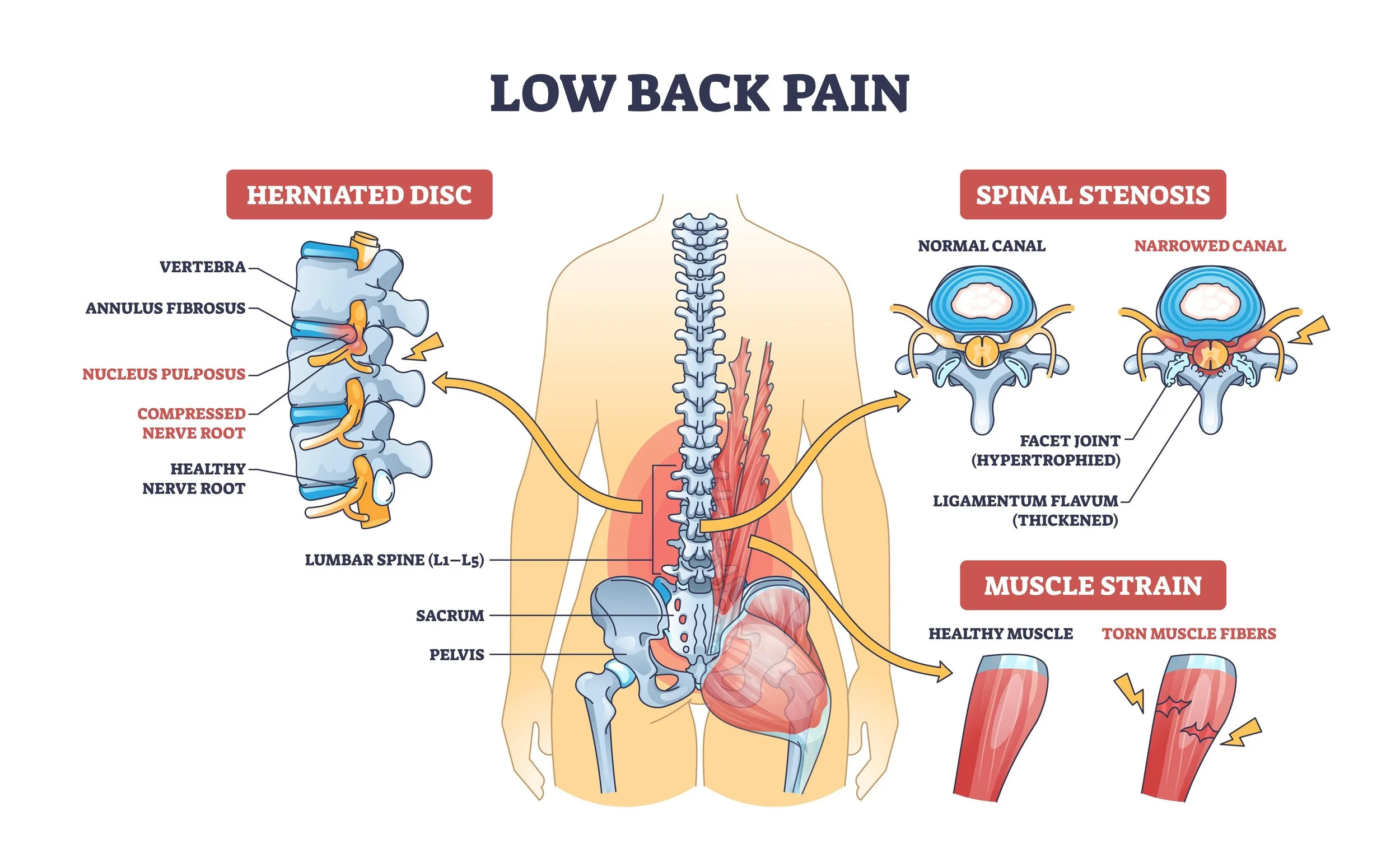
Do not be satisfied with a generic diagnosis of “spinal stenosis.” Find out which of the following causes of narrowing of the space between the spinal cord and the boney vertebrae applies to you.
Subluxation, or shifted vertebrae; i.e., they’re not stacked in alignment on top of each other.
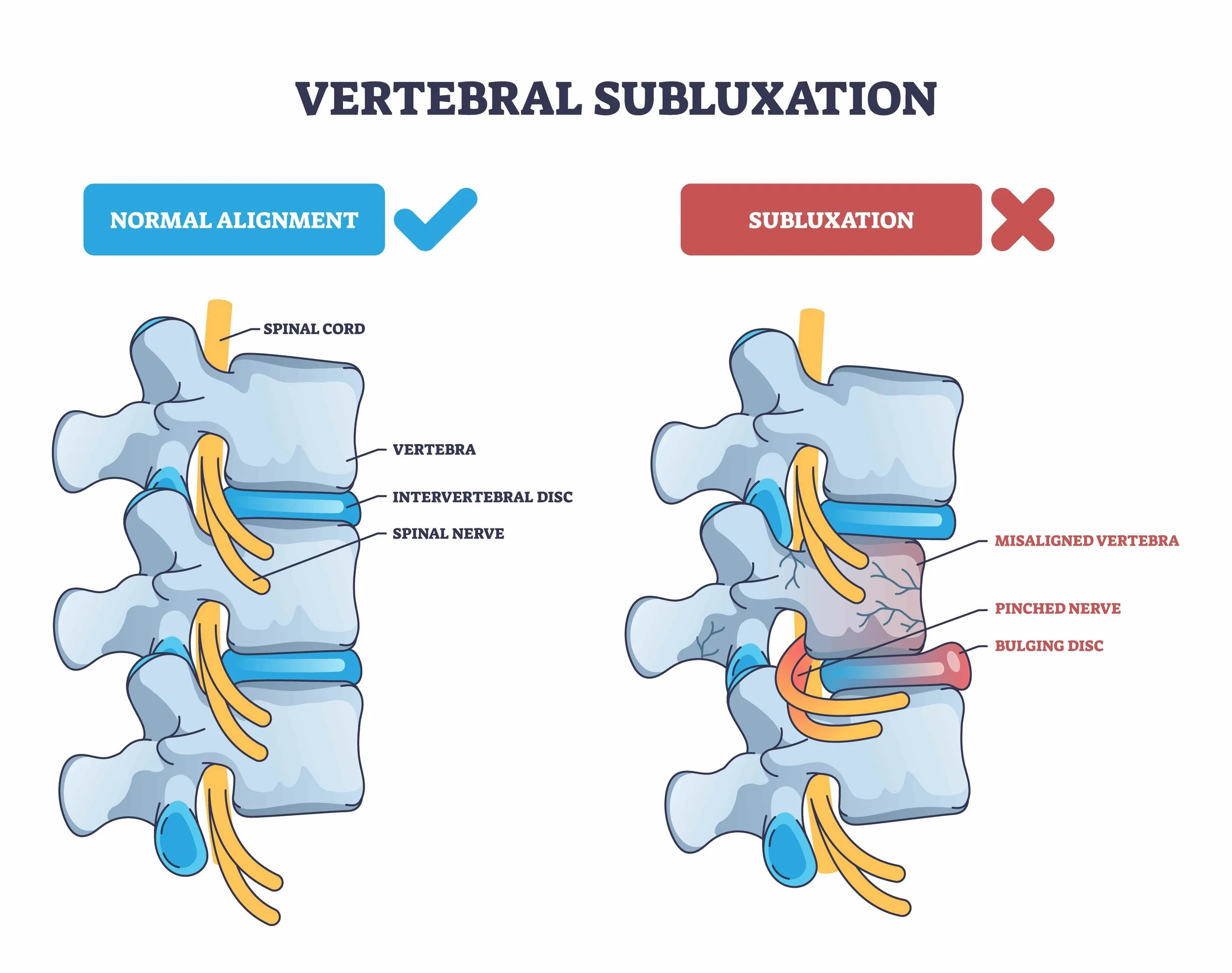
Bulging or Herniated Discs that protrude into the spinal cord and/or compress nerve roots where they exit the spinal cord.
Bone spurs (overgrowths of vertebral bone) that reduce the size of boney canal containing the spinal cord, compressing the cord or nerves.
Thickened Ligaments: The strong cords holding the spine together can stiffen and thicken over time.
Spinal Injuries: past fractures.
Congenital: born with a naturally smaller spinal canal.
All of the above causes can compress nerves where they exit the spinal cord. The result is chronic pain.
The years take a toll. Studies show that roughly 20% to 30% of adults over age 60 have evidence of lumbar spinal stenosis on MRI or CT scans.
Painless Spinal Stenosis. Of those with structural narrowing seen on scans, studies show that over 80% remain completely asymptomatic (no pain, numbness, or weakness.)
Has your specialist considered the possibility that, despite what the scans show, Spinal Stenosis is not causing your back pain? Scans have limitations…
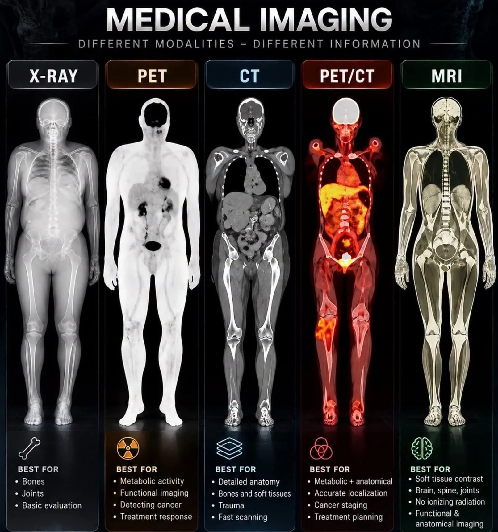
Physicians rely on various types of scans to diagnose a problem. Missing in the image above is Ultrasound imaging, the least expensive imaging option. It’s used for assessing soft tissues, organs, and blood flow without using radiation. For example, ultrasound can identify joint inflammation and tears in muscles, tendons, and ligaments.
Say the scan your physician ordered shows you do have spinal stenosis, and you do have back pain. Is spinal stenosis logically the cause of that back pain? Not necessarily…
Even if you have spinal stenosis (narrowing of the canal), you may have no compression of the nerve root where it exits the spinal cord.
Nerve compression is what causes the pain. The nerve may be compressed external to the spine.
After nerves exit the lumbar spine, and are external to the vertebrae, those long nerves wind their way through a lot of muscle and tissue. That is, nerve compression (and pain) may be caused by:
Option A: tight muscles or scar tissue in the lower back, or
Option B: tilted lumbar vertebrae due to pelvis misalignment.
Let’s discuss each in turn.
Option A. Tight Lumbar Muscles or Scar Tissue
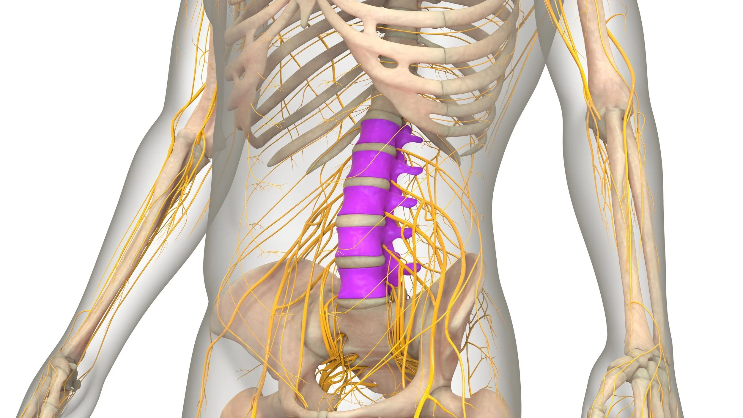
Due to the limits of technology, nerve compression per se is not visible on a scan. If a scan or palpation shows evidence of excess lumbar muscle tension, you can assume nerve compression. As you see above, there are a lot of nerves in that lower back!
Surgery will not resolve a lumbar nerve compression problem that has nothing to do with discs, subluxation (vertebra shifted out of alignment), and/or pressure on the spinal column. Physicians may instead recommend physiotherapy.
I can guarantee you that stretching does not release muscle knots and scar tissue compressing those lumbar nerves! Badly damaged tissue does not stretch. Instead, normal muscle fibers adjacent to the damage will rip, making things worse. Manual therapy techniques, acupuncture and dry-needling do address muscles locked in contractile tension, muscle knots and scar tissue. TENS is for nerve pain, and does not clear muscle damage.
Most therapists tend to target the area of pain, not the contracted, damaged muscles causing pelvis rotations, discussed below.
Option B. Tilted Lumbar Vertebrae
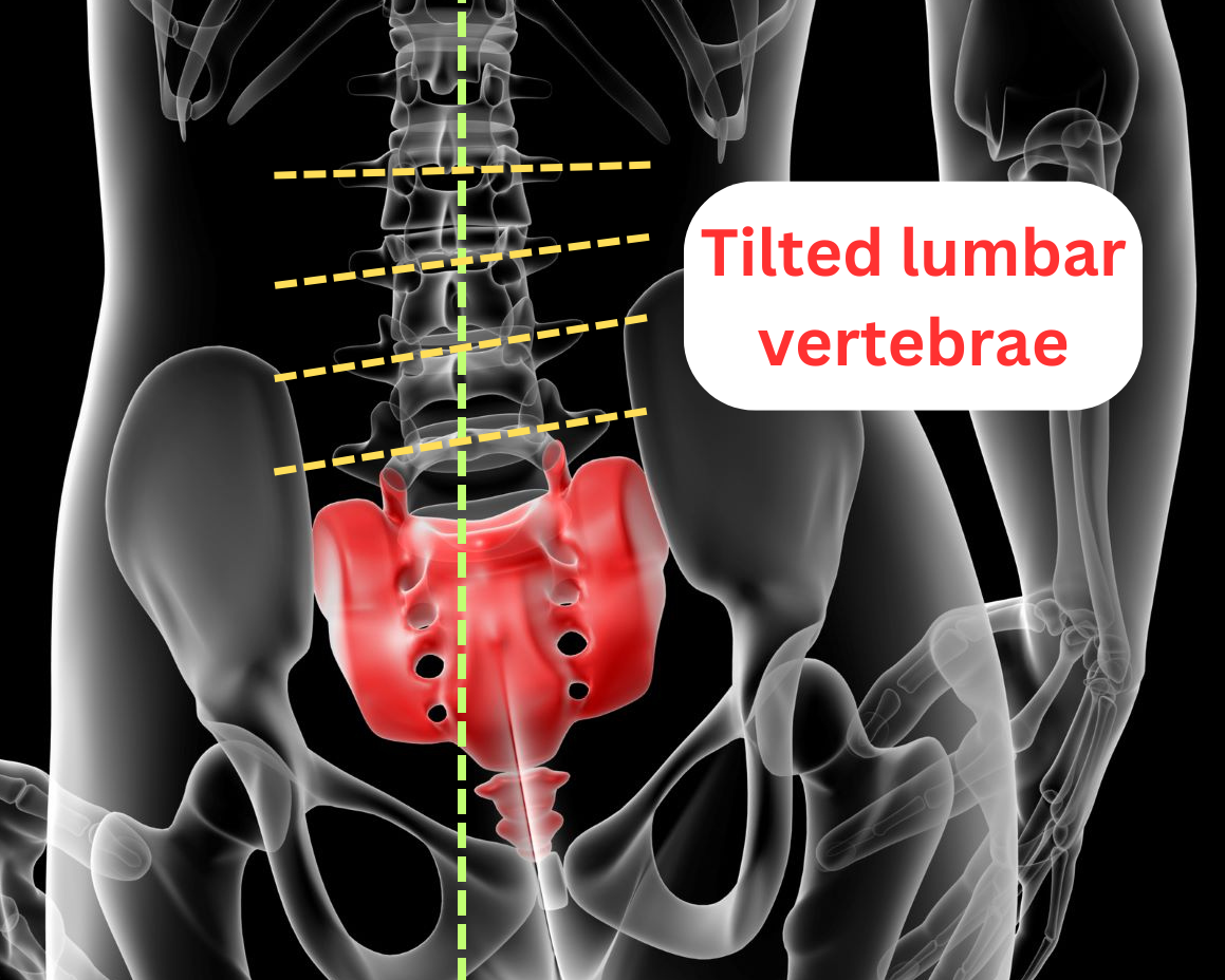
Tilted vertebrae are always caused by pelvis misalignment. For more on pelvic rotations, read my blog post Pelvis Rotations and Upslips .
Briefly, an anterior or posterior ilium (innominate) rotation causes:
Lordosis (X-ray lateral /side view)
Scoliosis (X-ray front and lateral views)
Misaligned vertebrae (X-ray front and lateral views):
-one or more vertebra shifted side/side or front/back, often by impact injuries, or
-one or more vertebra tilted into scoliosis by misaligned innominate (ilium) bones, as in the image above.
Tilted vertebrae cause many of the issues listed in scan reports; i.e.,
Facet bone osteoarthritis due to bones rubbing together,
Bone spurs/overgrowth,
Bulging or herniated discs.
Any of these can compress nerves.
When one side of the pelvis is higher than the other, clearly visible on an X-ray, back pain is likely caused by compression of nerves by tilted lumbar vertebrae transverse processes.
Back surgery will not correct lumbar spine or pelvis misalignment. As in this image below, post surgery the pelvis is clearly still misaligned, and the lordosis has not been corrected. Hip replacements are in this unfortunate patient’s future.
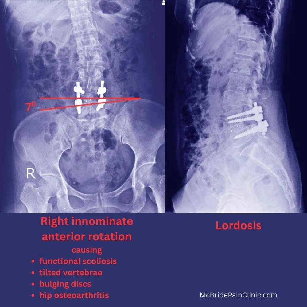
Back surgery would have been avoided if:
surgeons and radiologists were trained to recognize innominate (ilium) misalignment as a fundamental cause of back pain; and
the patient had been treated by a therapist trained in restoring pelvis and lumbar spine alignment.
Granted, that training is not easy to find, which is why I am teaching Biomechanics of Back, Hip and Knee Pain.
Case: Teenager With Chronic Back Pain
I included this case to illustrate how complex the causes of unrelenting pain can be. This teen had extensive muscle damage that pulled her skeleton out of alignment from neck to knee.
Nicole had fallen off her bike two years before seeing me. During those two long years her chronic pain was so bad that she could not sit for very long.
Before her parents found me, Nicole had seen many specialists. She’d had an X-ray and an MRI, both of which clearly showed a misaligned pelvis; i.e., the left ilium was significantly lower than the right, and that left ilium is clearly internally rotated. However, pelvis misalignment was not mentioned in the radiologist reports. They did mention disc bulges at L4, L5 and S1. Bulging discs are caused by tilted vertebrae (aka scoliosis) clearly visible on her X-ray.
A rheumatologist diagnosed her with Ankylosing Spondylitis. She’d been prescribed pain medication that gave her stomach ulcers.
A colonoscopy revealed her descending colon was blocked and it had to be “flushed out” twice. I had seen constipation like this before in other clients with an internally rotated left ilium that compressed the descending colon.
In the expensive and unsuccessful search for effective treatment, her parents took her for physiotherapy, chiropractic, osteopathy, acupuncture, and to a famous back specialist who recommended exercises and stretches.
Madeline’s Initial Assessment
Standing, Nicole’s left iliac crest was 1.5 inches lower than the right. Seated they were even. This indicated to me she had a double-ender problem — the cervical spine needed treatment as well. For those who have taken my workshop, the Front Line Test revealed weak left leg.
Palpation revealed significant muscle damage in her right lumbar and side from the bike accident. None of the therapists in two years of treatment had cleared this muscle tension. Her right piriformis was stretched, indicating compression of her sciatic nerve. No wonder she couldn’t sit for any length of time.
On her left side, erectors and lumbar muscles were contracted, but not torn in the accident. Her left quad was a solid block of contractile tension, plus she had deep, strong contractile tension the entire length of her left iliopsoas, both of which contributed to a left ilium anterior rotation. She had damage in her left hamstrings and muscles connecting to the Greater trochanter, typical of hamstring stretching to counter the left ilium anterior rotation.
Tight neck muscles pulled her left occiput inferior. She also had a torn left rotator cuff muscle and significant tension in her upper right back muscles.
Trapped Nerves
By the fifth session most of the tension in the larger back muscles was cleared and I was able to release muscle tension at the depth of the psoas attached to the left lumbar spine.
Addressing deep muscle tension is key to releasing compression of nerves exiting spinal vertebrae. Per Gil Hedley, two layers of psoas muscle attach to the lumber vertebral bodies and their transverse processes. A nerve adjacent to the spine passes vertically through that muscle. Pain from psoas muscle tears can be excruciating.
Over seven 75 minute sessions one summer I released most of the muscle tension from legs to jaw. Her pelvis was aligned. Her jaw and occiput were aligned. Her pain did not disappear because, against my advice, she continued to do the exercises recommended by the famous back specialist. Muscles need time to heal.
Scans Do Not Tell the Whole Story
Scans do not show the extent of muscle damage and contractile tension compressing nerves external to the vertebrae. Palpation, however, does.
Scans clearly show the extent of misalignment of the pelvis and lumbar spine.
Be aware most radiologists (at least in North America) are not taught to connect the dots between innominate rotations, lordosis, scoliosis and their effects; i.e., bulging or herniated discs, nerve compression by tilted transverse processes, and colon constriction. Maybe AI will make the connection for them…
WRAP UP
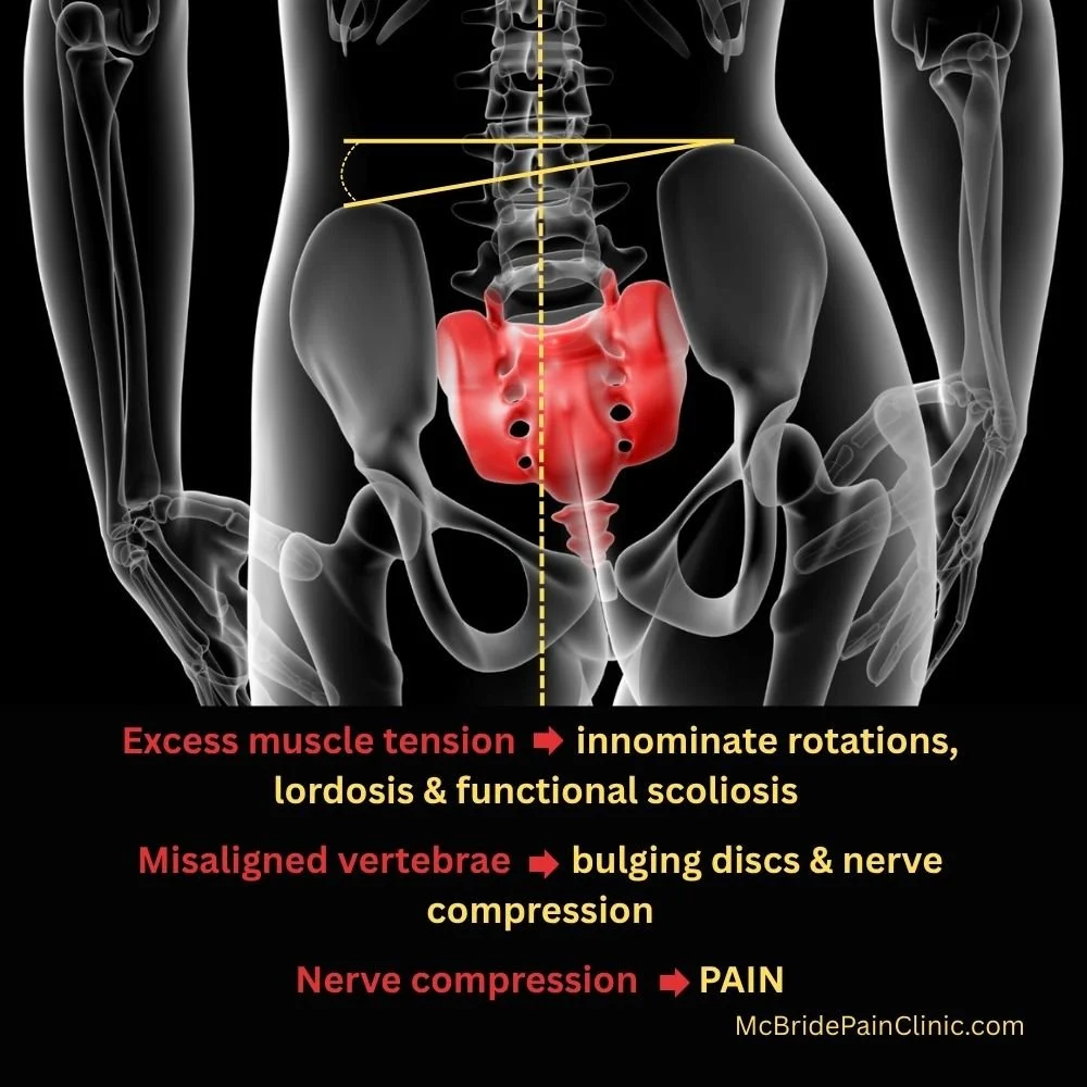
Surgeons do not correct lordosis nor pelvis innominate rotations when installing hardware in the spine.
If your pelvis and/or lumbar vertebrae are misaligned, be mindful that surgery may not eliminate nerve compression.
Post-spinal surgery, manual therapy cannot restore pelvic and lumbar alignment. It’s too late because the vertebrae are fixed in place.
Surgery should be a last resort. Book several sessions with a manual therapist trained in restoring myoskeletal alignment. In another post, I will provide qualifying questions you can ask to determine if the therapist has the expertise.
Author Bio: Madeline McBride, M.A.Sc., P. Eng., studied civil engineering at Queen’s University and the University of Waterloo. Those mechanics and structural design courses inform her ability to assess in 3D, and problem-solve how muscle tension pulls a client’s skeletal structure out of alignment. Her engineering background, combined with several manual training modalities, produced this neuromuscular therapist and Canadian expert in restoring pelvis, jaw and spinal alignment. Madeline is able to resolve recent ilium rotations and related back, hip, knee and neck pain within one to four sessions. Long-standing, complex issues may take longer due to the body’s entrained compensation. Madeline teaches Biomechanics of Back, Hip, and Knee Pain in-person and on-line. She teaches Jaw and Sphenoid Alignment in-person only. Register for a workshop or read her blog posts at www.McBridePainClinic.com